State-of-the-Art CT Scanner Offers Fast, Detailed Imaging
A state-of-the art medical imaging scanner at Albany Medical Center is offering patients of all ages a better, more comfortable experience, while also helping physicians make more accurate diagnoses by providing fast, highly detailed images of the heart, brain, cardiovascular system, and more.
The GE Revolution 256-slice CT scanner captures high resolution images very quickly, even if the patient’s heart rate is high—such as during a cardiac event—or if the patient has metal implants. It is most often used for cardiac imaging, stroke diagnosis, and detailed bone imaging.
“Our state-of-the-art imaging supports the best patient care,” said Dominic Zanello, manager of CT scan and MRI. “This 256-slice CT technology—the most advanced imaging capability available in the region—provides three dimensional cross-sections, allowing us to see what’s invisible to the naked eye, layer by layer, detail by detail.”
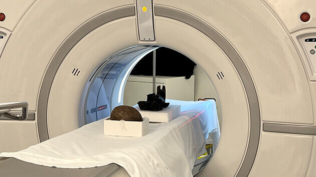
Recently, the technology’s advanced capabilities caught the attention of the New York State Museum, where experts were hoping to learn more about two paleontological artifacts. Experts visited the hospital after hours with a 70- to 80-million-year-old dinosaur egg and the skull of a giant beaver, an ice-age mammal that was as large as a modern black bear.
While no clear evidence of an embryo was seen inside the egg, a small cavity was detected and will be further analyzed. Robert Feranec, PhD, director of research and collections and curator of Pleistocene vertebrate paleontology at the New York State Museum, noted that the analyses could hold information about dinosaur reproduction and embryonic development for dinosaurs during the late Cretaceous Period and help scientists better understand the ecology of the extinct giant beaver and how it’s different from modern species.
“Our scientists are recognized as some of the best in their fields, but we can only see so much with the naked eye. There is much more we can learn from both the beaver skull and dinosaur egg, with these results acting as momentum pushing us closer to our answers,” Dr. Feranec said. “Collaborations like this are crucial to unlocking and preserving New York State’s past.”
Patients benefit from the scanner’s advanced capabilities daily. Sometimes called a “CAT scan”, a CT (computed tomography) scan uses a narrow x-ray beam that is rotated at a high rate of speed around the body to produce cross-sectional images (“slices”) that are processed by a computer to generate 3D images. Radiologists and other members of the health care team then use the scans to help diagnose disease or injury, as well as to plan medical, surgical, or radiation treatment.
Standard CT scans most often provide 64- or 128-slice images. With 256 slices, the Revolution scanner offers much more detailed information than conventional x-rays or even other CT scanners. The scanner’s speed also means patients are exposed to up to 82 percent less radiation. “It’s so fast that many patients, including kids, don’t require sedation,” added Zanello.
The Revolution can be used to visualize nearly any part of the body, from bones and joints to whole organs, including the brain, liver, kidneys, and pancreas. The design also allows the scanner to capture an image of the whole heart in a single beat for calcium scoring, coronary imaging, or a comprehensive cardiac assessment, with or without beta blockers.