Multiphoton Microscope Adds Speed, Clarity to Biomedical Research
Albany Medical College’s basic science researchers regularly use high-tech microscopes to study the mechanisms of disease—from how cancer cells move in the body, to potential new targets for treatment, to where and how antibodies bind to cells.
With the College’s newest microscope, the state-of-the-art Nikon AX R MP, installed last spring, the scientists are able to look even deeper into everything from proteins and molecules to arteries and tumors.
“Depth and resolution are two common problems with most microscopes, but the Nikon AX R MP has a tunable two-photon laser, allowing us to see larger structures in 3D, while maintaining high resolution quality,” explained Margarida Barroso, PhD, professor in the Department of Molecular and Cellular Physiology and co-director of the Imaging Core at Albany Medical College.
Accelerating Biomedical Research
Speed is also an issue with other, single photon microscopes, noted Dr. Barroso. To obtain an image of a larger biological structure like an artery or organ, most microscopes collect multiple images of specific sections, then tile them together, a process that can take three to four hours. The new Nikon microscope can produce the same image in about two minutes.
“Procedures that used to take hours can now be done in minutes,” said Arun Asif, PhD, a postdoctoral fellow in Dr. Barroso’s lab who has been using the microscope to visualize tumor structures in 3D.
“Imaging sessions that once stretched over a week can be finished in just a few hours,” he added. “This has changed how quickly we generate data, test new questions, and move projects forward.”
Dr. Asif has also used the microscope to track tumor cell invasion over time and to quantify collagen organization through second harmonic generation imaging (SHG), an advanced type of imaging that produces high resolution images.
“Before this microscope, most of our imaging had to be limited to thin sections of surface-level scans,” he said. “Now we can image deep inside tumors with high resolution, minimal photobleaching, and far better clarity.”
Viewing Larger Structures in 3D
Kevin Pumiglia, PhD, professor in the Department of Regenerative and Cancer Cell Biology, has also been “getting to know” the microscope and all its capabilities.
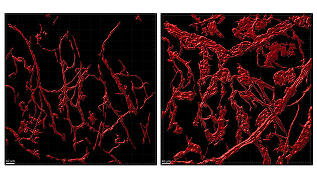
His lab studies the molecular mechanisms that cause vascular malformations—abnormal growths of blood vessels caused by either inherited or sporadic mutations—by modeling the mutations in complex cell cultures with multiple cell types, and in mice. With the new Nikon, they’re able to capture the abnormal blood vessels in 3D.
“Conventional microscopy presents multiple technical obstacles to imaging fields thicker than about 25 microns, but the two-photon microscope bypasses several of these limitations,” Dr. Pumiglia explained. “We can image deeper into tissues, much, much faster. This allows us to capture wider fields of view and with higher resolution than ever before.”
So far, they’ve used the microscope to attain higher-power images of blood vessels in both normal mouse ear skin and in that of a mouse that has a mutation in its endothelial cells (the cells that line the inside of blood vessels).
“We are imaging from the surface of the tissue down about 100 microns,” explained Dr. Pumiglia, who added that the images are then rendered to approximate a 3D representation. (A micron is one-thousandth of a millimeter.)
“This really allows us to see and appreciate the level of detail in the expansion of the endothelial cells and the overgrowth in the mutant ear skin,” said Dr. Pumiglia.
Eventually, he and his team hope to use the microscope’s live-imaging features to capture, over time, the emergence of vascular malformations on the ear skin of live mice. “If we can visualize in real-time the endothelial cells, blood flow, and more, we can learn what is going wrong with the mutated cells,” he added.
Advancing Biomedical Science in the Capital Region
Elsewhere within Albany Medical College, Bibhuti Mishra, PhD, assistant professor in the Department of Immunology and Microbial Disease, is using the Nikon microscope to better understand the pathogenesis of tuberculosis, and Alejandro P. Adam, PhD, associate professor in the Department of Molecular and Cellular Physiology, is using the microscope in his research on the role of endothelial cells in organ failure.
“So far, about ten Albany Medical College investigators have used the microscope, primarily for cancer, immunology, and inflammation research,” said Dr. Barroso.
But while it’s the only two-photon, two-laser microscope in the Capital Region, its use is not limited to Albany Medical College scientists.
Through an agreement with Rensselaer Polytechnic Institute, scientists at both institutions have access to specific equipment at each other’s institution, including the Nikon AX R MP.
“We’re also working to expand access and encourage participation from additional regional institutions and collaborators, including researchers from Wadsworth Center, the University at Albany, and industry,” said Dr. Barroso, who added that the microscope’s purchase and installation were supported by an NIH Shared Instrumentation (S10) grant and by Albany Medical College institutional funds.